
PAPER AT A GLANCE
Bioengineered immunocompetent preclinical trial-on-chip tool enables screening of CAR T cell therapy for leukaemia
Authors: Chao Ma, Huishu Wang, Lunan Liu, Ruiqi Chen, Nandana Mukherjee, Jie Tong, Shadab Kazmi, Xiangyi Fang, Matthew T. Witkowski, Iannis Aifantis, Saba Ghassemi*, Weiqiang Chen* (*corresponding authors)
Institutions: New York University (Tandon School of Engineering; Grossman School of Medicine; Perlmutter Cancer Center); University of Pennsylvania (Perelman School of Medicine; School of Engineering and Applied Science); University of Colorado Anschutz Medical Campus
Journal: Nature Biomedical Engineering 2025; 9(12): 2098–2114 · DOI: 10.1038/s41551-025-01428-2
Array used: RayBiotech Human Inflammation Antibody Array C3, membrane (cat. no. AAH-INF-3-8)
License: Open access, CC BY 4.0
In one sentence: the authors built a vascularized, immune-competent “leukaemia bone marrow on a chip” and showed that it can reproduce remission, resistance and relapse after anti-CD19 CAR T cell therapy — and rank CAR T cell products from different donors, designs and manufacturing protocols before they reach patients. A membrane cytokine array, the RayBiotech Human Inflammation Antibody Array C3, was the read-out they used again and again to see how each CAR T cell product talked to the leukaemia niche.
Why this question matters
CAR T cell therapy has transformed relapsed and refractory B-cell leukaemia, but outcomes are hard to predict. The paper notes that nearly half of patients in some trials eventually relapse, and there are still no molecular biomarkers that reliably forecast who will respond. Preclinical testing is part of the problem. Two-dimensional co-cultures and tumour spheroids can show that CAR T cells kill target cells, but they cannot show T cells leaving blood vessels, migrating through bone marrow tissue, or interacting with the stromal and immune cells around the tumour. Mouse models add a body, but immunocompetent models with a human immune system are slow to build and still not human.
What the team built
The device is a PDMS microfluidic chip with three concentric compartments that mirror the anatomy of leukaemic bone marrow: a central sinus, a medullary cavity and an outer endosteal region. Each compartment is filled with fibrin gel seeded with primary human cells — vascular cells, mesenchymal stem cells, osteoblasts, fibroblasts and bone marrow mononuclear cells — plus GFP-labelled B-ALL leukaemia cells. Over a week the vascular cells self-assemble into perfusable vessels, so CAR T cells can be infused through the “blood” side and tracked as they extravasate and hunt leukaemia cells in real time by imaging.
According to the authors, a chip can be assembled in about half a day and supports at least 14 days of CAR T cell monitoring, which compares favourably with the months needed to establish humanized immunocompetent animal models.
Key findings
- The chip reproduces effective therapy. 10,000 second-generation anti-CD19 4-1BBζ CAR T cells per chip killed about 70% of leukaemia cells by day 3 and more than 99% by about day 7, while non-transduced (“mock”) T cells did not control the leukaemia.
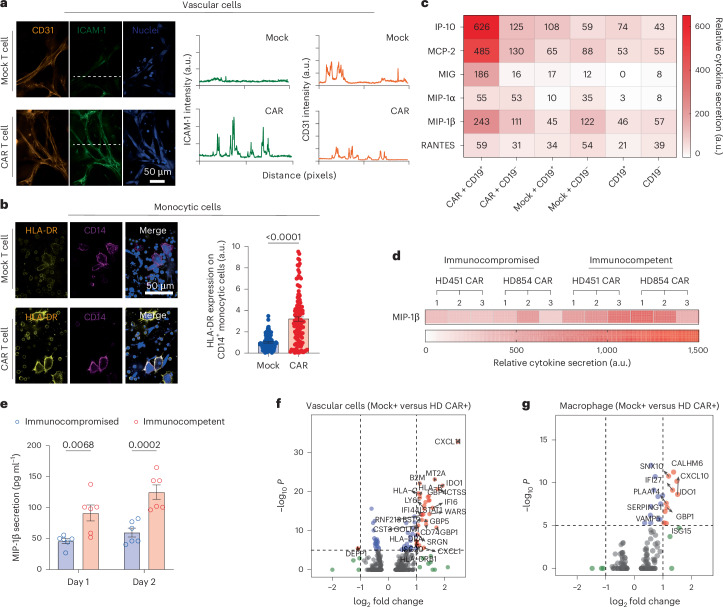
- CAR T cells switch on the whole niche. After infusion, vascular cells increased ICAM-1 and CD14+ monocytic cells increased HLA-DR. Cytokine array profiling showed a broad surge of chemokines and inflammatory cytokines — for example MIP-1β, RANTES and MCP-2 — only when CAR T cells met CD19+ leukaemia cells, not with mock T cells or CD19− targets. Chips that contained bone marrow mononuclear cells (immunocompetent) secreted more MIP-1β than chips without them, a difference the authors confirmed by ELISA.
- Remission, resistance and relapse can all be modelled. A low dose (2,500 CAR T cells) mostly failed, mimicking refractory disease. Spiking in 1–5% CD19− leukaemia cells produced initial clearance followed by outgrowth of the antigen-negative clone, mimicking antigen-loss relapse. IFN-γ secretion was highest in the remission scenario, and the authors compared these on-chip response patterns with tumour-burden data from 209 patients treated in their earlier clinical study.
- Tumour burden and bridging therapy matter. High leukaemia burden blunted the response, whereas 24 h of vincristine “bridging chemotherapy” before CAR T cell infusion improved it — and CAR T cells spread further through the chip.
- Products can be ranked before the clinic. The chip separated CAR designs (4-1BBζ CAR T cells secreted more of the TH2 cytokine IL-13, while CD28ζ and ICOSζ CARs secreted more IL-10), manufacturing protocols (CAR T cells expanded for 3 days outperformed 9-day products at low doses), patient-derived products (only one of four patient products achieved on-chip remission), and a fourth-generation CAR armoured with IL-18, which boosted cytokine output and response.
How the cytokine antibody array was used
The methods section describes a straightforward workflow that many labs running co-culture or organ-on-chip experiments could copy:
- Sample: supernatants pooled from 2–4 chips at defined time points after CAR T cell or mock T cell infusion.
- Clean-up: centrifugation at 2,000 × g for 20 min at 4 °C to remove debris; samples were run fresh or stored at −20 °C.
- Assay: overnight incubation of supernatant on the array membranes, overnight incubation with the biotinylated detection-antibody cocktail at 4 °C, then HRP-streptavidin and chemiluminescent detection, following the kit protocol.
- Read-out: qualitative, relative profiles of 40 inflammation-related proteins per membrane, used to compare conditions side by side.
- Confirmation: targets that stood out on the array — MIP-1β, IFN-γ and granzyme B — were then measured quantitatively by ELISA.
The array appears throughout the paper: niche crosstalk (Fig. 3), CAR designs (Extended Data Fig. 4), 3-day versus 9-day manufacturing (Extended Data Fig. 5), patient-derived products (Extended Data Fig. 6) and the IL-18-armoured CAR (Fig. 5). The authors also grouped the cytokines into five functional classes — effector, stimulatory, chemoattractive, inflammatory and regulatory — and combined them into a cytokine secretion index to rank products.
Why a membrane array fits this kind of study: chip supernatant volumes are small, the biology is exploratory (you do not know in advance which of dozens of cytokines will move), and many conditions need to be compared. A 40-target screen per sample, followed by ELISA for the few targets that change, is an efficient division of labour.
Strengths and limitations — a journal-club view
Strengths
- Human cells in a vascularized, compartmentalized architecture, so trafficking and extravasation can be watched, not inferred.
- Several clinically recognisable scenarios — remission, resistance, antigen-loss relapse, high tumour burden and bridging therapy — modelled in a single platform.
- Multidimensional read-outs: live imaging, flow cytometry, single-cell RNA-seq and cytokine profiling.
Limitations the authors acknowledge, and points to keep in mind
- The system is allogeneic; the authors observed possible graft-versus-host effects, such as fewer CD45+ cells and damaged vessels.
- Correlation between on-chip results and clinical outcome in the same patients remains to be established; only a small number of patient-derived products were tested.
- Some array comparisons rest on two experiments (n = 2), and membrane arrays give relative rather than absolute values — which is why the ELISA confirmations matter.
- Acquired antigen mutation and other resistance routes were not modelled.
Questions to discuss in your lab
- Would a cytokine secretion index measured on-chip predict cytokine release syndrome risk as well as efficacy?
- Which read-outs would you add to separate T cell exhaustion from insufficient expansion in the “resistance” scenario?
- How would an autologous (fully patient-matched) chip change the interpretation of the cytokine data?
Running a similar cytokine screen
The array used in this study is the RayBiotech Human Inflammation Antibody Array C3 (AAH-INF-3), a semi-quantitative membrane array for 40 human inflammation-related proteins read on a standard chemiluminescence imager. For chemokine-focused questions, see the Human Chemokine Array C1; to choose between screening and quantification, see RayBiotech antibody arrays and ELISA kits. Request a quote for any of these products.
Reference
Ma C, Wang H, Liu L, Chen R, Mukherjee N, Tong J, Kazmi S, Fang X, Witkowski MT, Aifantis I, Ghassemi S, Chen W. Bioengineered immunocompetent preclinical trial-on-chip tool enables screening of CAR T cell therapy for leukaemia. Nature Biomedical Engineering. 2025;9(12):2098–2114. doi:10.1038/s41551-025-01428-2. Publisher · PubMed Central
This summary is an independent commentary by Changyu Bio written for researchers; it is not affiliated with or endorsed by the authors or the journal. All products mentioned are for research use only.