
Searches for running mRNA on an agarose gel usually come from one of two benches: a researcher checking an enriched poly(A)+ mRNA sample, or someone performing quality control on an in vitro transcribed (IVT) transcript. Both often end up surprised, because mRNA does not look like total RNA on a gel — and reading it with “total-RNA eyes” is the single most common mistake on this topic.
This article explains what to expect for each situation, featuring real lab data for direct comparison.
First: The Picture You Should NOT Expect
If your gel shows two sharp, bright bands with the upper band roughly twice as intense as the lower one, you are looking at total RNA, not mRNA. Those are the 28S and 18S ribosomal RNAs (rRNA), and they only appear when ribosomal RNA is still present in the sample.
Here is a reference gel from our lab — total RNA extracted using the Changyu Bio RNApure Total RNA Kit, run on a 1% agarose gel:
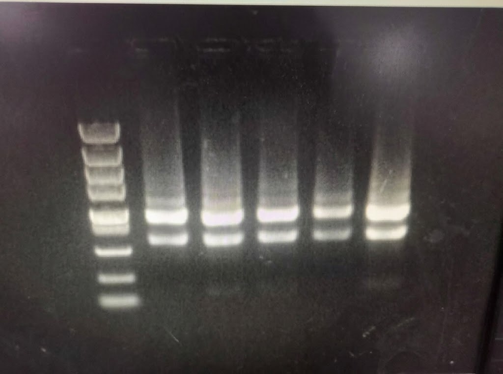
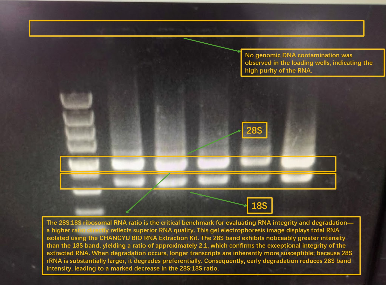
(For our complete guide to interpreting total RNA gels — including degradation smears, genomic DNA contamination, and bright wells — see our dedicated protocol on checking RNA quality by agarose gel electrophoresis.)
Situation 1: Poly(A)+ mRNA — A Continuous Smear Is Expected
Oligo(dT) or bead-based enrichment removes ribosomal RNA. Consequently, a high-quality poly(A)+ sample should show no distinct 28S or 18S bands. Instead, it migrates as a continuous smear spanning roughly 0.5–10 kb, typically peaking around 1–2.5 kb depending on the organism. This represents the natural size distribution of cellular mRNAs.
Do not mistake this smear for degradation. In poly(A)+ mRNA, the smear is the expected signal. What you should actually watch out for:
- Downward-shifted smear: A distribution skewed heavily toward the bottom of the lane (most signal below ~1 kb) indicates degraded or fragmented mRNA.
- High-molecular-weight band: A distinct band trapped near or just below the well indicates genomic DNA (gDNA) carryover.
- No signal: Suggests low recovery or sample loss during enrichment; re-quantify concentration (e.g., via NanoDrop or Qubit) before troubleshooting the gel.
Situation 2: IVT mRNA — One Sharp, Discrete Band
For synthetic, in vitro transcribed mRNA of a defined sequence, the objective is verifying transcript integrity: the full-length product should dominate, with minimal truncated material. (Note that capping and poly(A) tail quality are assessed by other methods such as HPLC or mass spectrometry — not by gel.)
- Run a denaturing gel: Use formaldehyde/MOPS or glyoxal/DMSO conditions to eliminate secondary structures. Without denaturation, RNA secondary structures cause unpredictable migration, preventing accurate molecular weight sizing.
- Expected profile: Look for a single, sharp band corresponding to your target transcript size. Smeared signal below this band reflects prematurely terminated (truncated) products; material stuck in the well indicates aggregation.
- Use an appropriate ladder: A single-stranded RNA ladder covering your target size range is essential for confident sizing.
(A quick non-denaturing run can serve as a preliminary sanity check to confirm product presence, but rigorous sizing requires denaturing conditions.)
Recommended Practical Protocol
- Gel Matrix: 1.0–1.2% agarose in 1× MOPS buffer containing ~2.2 M formaldehyde (for denaturing runs); standard 1× TAE or 0.5× TBE buffer suffices for non-denaturing quick checks.
- Sample Denaturation: Mix with formamide/formaldehyde RNA loading buffer, incubate at 65–70 °C for 10 minutes, and immediately chill on ice prior to loading.
- Electrophoresis: Run at ~5 V/cm until the tracking dye migrates approximately two-thirds down the gel.
- Staining: Use sensitive, post-staining RNA fluorophores (such as SYBR Gold, GelRed, or SYBR Safe) or standard ethidium bromide post-stain.
- RNase Precautions: Maintain dedicated RNase-free tanks, clean combs with 0.1 M NaOH or RNase decontamination solutions, and use DEPC-treated or nuclease-free water.
Summary
- Two sharp bands (28S ≈ 2× 18S): intact total RNA.
- 0.5–10 kb continuous smear: expected profile for enriched poly(A)+ mRNA.
- Single crisp band at expected MW: high-quality IVT mRNA.
- Denaturing conditions: required whenever exact size determination is critical.
- Degradation indicator: a downward shift of the signal towards low molecular weights (<1 kb).
Looking to ensure optimal starting material? Explore our RNApure Total RNA Kit and complete RNA isolation portfolio. For tissue or cell samples that will travel or wait before processing, RNAfixer stabilizes RNA at the point of collection.
All products are For Research Use Only (RUO). Not for use in diagnostic, therapeutic, or clinical procedures.
More questions
- Can I use the same gel to check total RNA? Yes — for total RNA you read the 28S and 18S bands rather than a smear. The RNA agarose gel electrophoresis guide covers band reading, degradation and DNA contamination.
Related gel and RNA quality guides
- What degraded RNA looks like on a gel
- 28S:18S rRNA ratio and band sizes
- Reading RNA integrity on agarose gel
- How to run RNA on a standard agarose gel
- RNA gel troubleshooting: extra bands, smears and odd sizes
- Genomic DNA quality on agarose gel
- A260/A280 for RNA: the complete guide
Frequently asked questions
What does mRNA look like on an agarose gel?
Poly(A)+ mRNA enriched by oligo(dT) or bead-based methods shows no distinct 28S or 18S bands. Instead it appears as a continuous smear spanning roughly 0.5–10 kb, typically peaking around 1–2.5 kb depending on the organism. This smear is the expected signal, not a sign of degradation.
Why does my mRNA gel not show 28S and 18S bands?
Sharp 28S and 18S bands represent ribosomal RNA. Enrichment removes most rRNA from poly(A)+ samples, so those bands are not expected. They only appear when rRNA is still present, which is why a total RNA gel and an mRNA gel look different.
How do I run in vitro transcribed mRNA on a denaturing gel?
Use 1.0–1.2% agarose in 1× MOPS buffer containing about 2.2 M formaldehyde. Mix samples with formamide/formaldehyde RNA loading buffer, heat at 65–70 °C for 10 minutes and chill on ice before loading. Run at about 5 V/cm and use a single-stranded RNA ladder for sizing.
What does a degraded mRNA sample look like on a gel?
A downward-shifted smear, with most signal below about 1 kb, indicates degraded or fragmented mRNA. A distinct band near or just below the well indicates genomic DNA carryover, and no signal suggests low recovery during enrichment.